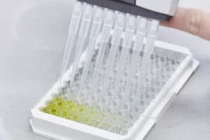
Sandwich ELISA test
Introduction
An immunoassay is a biochemical test measuring the concentration of a protein in a biological fluid. The assay takes advantage of the specific binding of an antibody to its antigen.
There are different types of immunoassays, this protocol contains information about a Sandwich ELISA (Enzyme Linked Immuno Sorbents Assay). In Sandwich immunoassay, also referred to as the “Non-competitive ELISA,” antigen is bound to the antibody site and a labeled antibody is bound to the antigen.
The amount of antigen on the site is measured. The results of the non-competitive ELISA method will be directly proportional to the concentration of the antigen. This is because labeled antibody will not bind if the antigen is not present in the unknown sample.
Procedure
- Before the assay, both antibody preparations should be purified and albumin-free. The detection antibody must be labeled.

Coating with capture antibody
2. Coat the wells by adding approximately 100 µl of coating antibody solution to each well. The amount of antibody used will depend on the individual assay, optimize the buffer and the coating concentration (1-10 µg/well).
3. Aspirate the coating solution.
Blocking
4. The remaining sites for protein binding on the microtiter plate must be saturated by incubating with blocking buffer. Fill the wells with 200 µl blocking buffer.
5. Cover the plate with adhesive plastic and incubate for 60-90 minutes at room temperature or overnight at 4°C.
6. Wash wells four times with wash solution.
Sample incubation
7. Add 100 µl of the diluted samples, standards and controls to the wells. All dilutions should be done in the dilution buffer.
8. Cover the plate with adhesive plastic and incubate for 60-90 minutes at 37°C.
9. Wash the plate four times with wash solution.
Incubation with primary antibody
10. Add 100 µl of the primary antibody. The amount to be added can be determined in preliminary experiments. For accurate quantification, the primary antibody should be used in excess. All dilutions should be done in the dilution buffer.
11. Cover the plate with adhesive plastic and incubate for 60-90 minutes at room temperature or at -20°C.
12. Wash four times with wash solution.
Incubation with the secondary antibody (or Streptavidine-HRP in case of biotinylated primary antibody)
13. Add 100 µl of the labeled secondary antibody. The amount to be added can be determined in preliminary experiments. For accurate quantification, the labeled secondary antibody should be used in excess. All dilutions should be done in the dilution buffer.
14. Cover the plate with adhesive plastic and incubate for 60-90 minutes at room temperature or at -20°C .
15. Wash four times with wash solution.
Substrate incubation
16. Add substrate as indicated by manufacturer.
17. After suggested incubation time has elapsed, add the stop solution to each well.
18. Optical densities at target wavelengths can be measured on an ELISA reader within thirty minutes after adding stop solution.
Analysis of the data
19. Calculate the average absorbance values for each set of duplicate standards, samples and controls.
20. If individual absorbance values differ by more than 15% from the corresponding mean value, the result is considered suspect and the sample should be re-assayed.
21. Create a standard curve by reducing the data using computer software capable of generating a good curve fit.
22. If the samples have been diluted, the concentration determined from the standard-curve must be multiplied by the dilution factor.
Technical specifications
Materials / reagents
- Coating antibody (1-10 µg/ml in coating buffer)
- Samples, standards and controls
- Primary antibody (unlabeled or biotinylated)
- Enzyme labeled secondary antibody
- Substrate
- Coating buffer:
0.15 M sodium carbonate, 0.35 M sodium bicarbonate, pH 9.6 (Carbonate Coating Buffer)
• 3.18 g sodium carbonate (Na2CO3)
• 5.86 g sodium bicarbonate (NaHCO3)
• Fill up to 200 ml with distilled water
• Adjust pH to 9.6 with hydrochloric acid Phosphate Buffered Saline (PBS), pH7.4
• 0,086 M disodium hydrogen phosphate (Na2HPO4)
• 0,020 M monopotassium phosphate (KH2PO4)
• 3,08 M sodium chloride
Blocking buffer:
PBS, 1% BSA
• 500 ml PBS
• 5 g BSA
Wash solution:
PBS, 0.05% Tween-20
• 400 ml PBS
• 2 ml Tween-20
• Fill up with to 4 L with distilled water
Dilution buffer:
PBS, 0.05% Tween-20, 0.1% BSA - 500 ml PBS
- 0.25 ml Tween-20
- 0.5 g BSA
PBS, 0,1%BSA
• 500 ml PBS
• 0.5 g BSA
Stop Solution; 2 % oxalic Acid
Safety
- Under no circumstances shall Hbt be liable for any damage arising from the use of this protocol. User should be trained and be familiar with the test procedure.
- Samples of tissue, serum or blood origin should be handled to guidelines for prevention of transmission of blood borne diseases.
- Some enzyme substrates are considered hazardous, due to potential carcinogenicity. Handle with care and refer to Material Safety Data Sheets for proper handling precautions.
- Wear appropriate protective clothing, gloves, and eyewear necessary to avoid any accidental contact with reagents.
- Use extreme caution, …. is a known carcinogen.
- Reagents which contain preservatives may be toxic if ingested, inhaled, or in contact with skin.
Notes
- Please note that this is a general protocol. Optimal dilutions for the primary and secondary antibodies, cells preparation, controls, as well as incubation times will need to be determined empirically and may require extensive titration. Ideally, one would use the antibodies as recommended in the product data sheet.
- The appropriate negative and positive controls should be included in every trial.
- For accurate quantitative results, always compare signal of unknown samples against those of a standard curve. Standards (duplicates or triplicates) and blank must be run with each plate to ensure accuracy.
- When making buffer(s) fresh before you start, there is no need to add sodium azide to the buffer.
- Do not include sodium azide in buffers or wash solutions if an HRP-labeled antibody/conjugate will be used for detection.
- Bring all reagents to room temperature before use.
- Mix all reagents well before use.
- Be precise and accurate during pipetting. Poor results may be the result of the following factors:
- a. The pipette-tip(s) are not fastened tightly to the pipette. Tips on multi-channel pipette should be checked individually to make sure that they are securely fitted.
- b. The pipette is filled too quickly; be aware of air bubbles in the pipette tip which may be formed when the liquid is drawn up too fast. (10 l of air instead of sample gives a variation of 10%!)
- c. Droplets clinging to the outside of the pipette tip.
- d. The pipette tip is emptied too quickly. To reduce variation during the pipetting steps, work at an uniform pace – one that minimizes the time between pipetting the first and last wells but which does not introduce errors due to hasty pipetting. Also, the micropipettes should be checked periodically for proper function and calibration.
- Use the incubation times and temperatures as specified in the protocol.
- Check plate for presence of air bubbles and make sure that all are removed (without removing any solution), prior to reading the plate. The spectrophotometer wavelength should be set accurately to the optimum wavelength.
- We recommend that disposable plastic tubes are employed when using TMB as substrate.
- Incubate plates uniformly in case of 37°C incubation. Plates should be placed directly on a prewarmed surface for incubation at 37°C. Use a humidified incubator to reduce drying effects in the outside wells (“edge effects”) or place a wet tissue or blotter paper under the plates in non-humidified incubators. Avoid opening the incubator frequently during the incubation period. Do not stack plates.
We are glad to support you!
Take advantage of our dedicated support team for any technical assistance you need while using our products or considering them for your research needs.