SAP, Mouse, pAb
Quantity:
100 µg
Catalog #:
HP8039
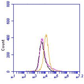
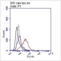
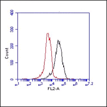
The polyclonal antibody recognizes serum amyloid P (SAP). Serum amyloid P component (SAP) belongs to a highly conserved superfamily of calcium-dependent ligand binding and lectin (carbohydrate binding) proteins. Due to the pentameric structure these proteins are also called pentraxins. SAP is 25kDa in size. C-reactive protein (CRP) and SAP are well-charactarized short pentraxins, which are produced in the liver in response to inflammatory mediators. Pentraxin 3 (PTX3) is the prototypic long pentraxin. Both SAP and CRP are evolutionary conserved in all vertebrates and found in distant invertebrates such as the horseshoe crab (Limulus polyphemus). They share 51% residue sequence identity and 66% homology.
SAP is highly resistant to proteolysis, especially when it forms complexes with calcium-dependent ligands. SAP avidly binds to macromolecular ligands, such as nucleosomal DNA, glycosaminoglycans and amyloid fibrils. When aggregated it can bind C1q and activate the classical complement pathway.
Human SAP is not an acute phase reactant following acute stimuli, this in contrast to mouse SAP and human CRP. In mouse, SAP levels increase significantly 24 hours after challenge with lipopolysaccharide.
SAP is a normal plasma constituent that is present in cerebrospinal fluid (CSF), in the pathognomonic lesions of Alzheimer's disease (AD), cerebrovascular and intracerebral Aβ amyloid plaques and neurofibrillary tangles. This is a result of its binding to amyloid fibrils and to paired helical filaments, respectively. The protein itself may also be directly neurocytotoxic. SAP contributes significantly to the pathogenesis of amyloidosis. The mechanism of its participation is not yet known and importantly may differ between species.
Product type | Polyclonal antibodies |
---|---|
Quantity | 100 µg |
Formulation | 1 ml (100 µg/ml) 0.2 µm filtered antibody solution in PBS, containing 0.1% bovine serum albumin and 0.02% sodium azide. |
Application | Frozen sections, Immuno assays, Immuno precipitation, Paraffin sections, Western blot |
Use | For immunohistochemistry, and Western blotting, dilutions to be used depend on detection system applied. It is recommended that users test the reagent and determine their own optimal dilutions. The typical starting working dilution is 1:50. For functional studies, in vitro dilutions have to be optimized in user's experimental setting. |
Immunogen | Purified natural mouse SAP |
Isotype | Sheep Ig |
Species | Mouse |
Storage and stability | Product should be stored at 4 °C. Under recommended storage conditions, product is stable for at least one year. The exact expiry date is indicated on the label. |
Precautions | For research use only. Not for use in or on humans or animals or for diagnostics. It is the responsibility of the user to comply with all local/state and federal rules in the use of this product. Hycult Biotech is not responsible for any patent infringements that might result from the use or derivation of this product. |
Disease | Autoimmunity |
Application: | Frozen sections, Immuno assays, Immuno precipitation, Paraffin sections, Western blot |
---|---|
Use: | For immunohistochemistry, and Western blotting, dilutions to be used depend on detection system applied. It is recommended that users test the reagent and determine their own optimal dilutions. The typical starting working dilution is 1:50. For functional studies, in vitro dilutions have to be optimized in user's experimental setting. |