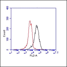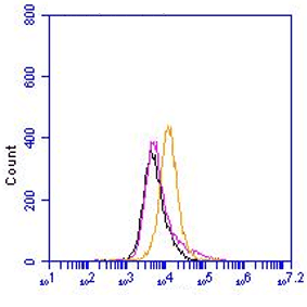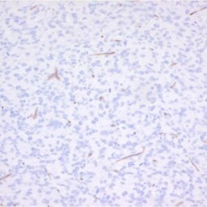

Monoclonal antibody PR3G-2 reacts with human proteinase 3 (PR3), a 30 kDa protein. PR3 is a major antigen recognized by autoantibodies directed against cytoplasmic proteins of neutrophilic granulocytes and monocytes (called anti-neutrophil cytoplasmic autoantibodies (ANCA)). ANCA are able to activate primed neutrophils to produce oxygen radicals and release lytic enzymes, including PR3. Proteinase 3 (PR3) was identified as the target antigen of ANCA in Wegener’s granulomatosis (WG). ANCA directed against PR3 (PR3-ANCA) can interfere with the binding of PR3 to its physiological inhibitor alpha1-antitrypsin (alpha1-AT) and with the proteolytic activity of PR3. At the site of inflammation, PR3 can cleave the PR3-ANCA complex between these inhibiting ANCA and PR3 itself, leaving active PR3. Autoantibodies to PR3 are potent activators of the 5-lipoxygenase pathway in primed human neutrophils. Extracellular free arachidonic acid, as present at an inflammatory focus, synergizes with such autoantibodies to evoke full-blown lipid mediator generation, granule secretion and respiratory burst. Proteinase 3 (PR3) is a neutral serine proteinase, which is localized in the azurophilic granules of neutrophils and in granules of monocytes and can be detected in the membrane of secretory vesicles. PR3 degrades a number of extracellular matrix proteins such as elastin and inactivates human C1 inhibitor. Membrane-associated PR3 is also able to activate caspase-3 without triggering apoptosis of neutrophils, which is possibly a neutrophil survival mechanism. In addition, PR3 is involved in myeloid differentiation and is, therefore, also called myeloblastin. The monoclonal antibody PR3G-2 was produced by immunization of mice with a crude granule extract.

 Powered by Bioz
Related products
-
View product €139.00 €333.00Price range: €139.00 through €333.00
-
View product €139.00 €9,220.00Price range: €139.00 through €9,220.00
-
View product €139.00 €431.00Price range: €139.00 through €431.00
-
View product €139.00 €1,294.00Price range: €139.00 through €1,294.00
-
View product €139.00 €383.00Price range: €139.00 through €383.00
-
View product €139.00 €333.00Price range: €139.00 through €333.00
-
View product €139.00