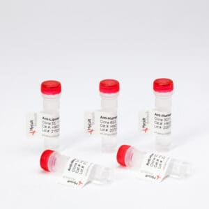
Custom concentrations and volumes possible
VAP-1, Mouse, mAb 7-88
€139.00 €588.00Price range: €139.00 through €588.00
The monoclonal antibody 7-88 recognizes mouse Vascular Adhesion Protein-1 (VAP-1) which- is a glycosylated homodimeric membrane protein consisting of two 90 kDa subunits connected by disulfide bonds. It contains a short N-terminal cytoplasmic tail, a single membrane-spanning domain and a large extracellular part. A soluble form of VAP-1 (sVAP-1) has been described, which presumably results from the proteolytic cleavage of membrane-bound VAP-1. Structurally VAP-1 belongs to enzymes called semicarbamizide-sensitive amine oxidases, which contain copper as a cofactor. These enzymes deaminate primary amines in a reaction producing hydrogen peroxide, aldehyde, and ammonia.
VAP-1 is expressed in endothelial cells, smooth muscle cells, adipocytes, and in follicular dendritic cells. In endothelial cells the majority of VAP-1 is stored within intracellular granules and translocated to the surface upon inflammation where it regulates leukocyte tissue infiltration. Furthermore, the end-products formed by VAP-1 can also regulate leukocyte migration by signaling effects, have insulin-like effects in energy metabolism, and can cause vascular damage by direct cytotoxicity.
In- white adipose tissue of obese and diabetic db-/- mice increased expression of VAP-1 has been observed suggesting that it contributes- to the arthrosclerosis and vascular dysfunction observed in these diseases. Moreover, inhibition of VAP-1reduced the accumulation of myeloid cells into tumors and attenuates tumor growth.
The monoclonal antibody 7-88 inhibits migration of granulocytes and monocytes in acute models of inflammation.
IHC-F:- Tissue sections were fixed in acetone and incubated with antibody 7-88 for 20 minutes at room temperature. . As negative control an irrelevant isotype-matched antibody was used (Ref.2).
FS: Antibody 7-88 (200 µm) was intravenously injected which resulted in the inhibition of leukocyte trafficking in inflamed peritoneum.- (Ref.2).

