How to Analyze Complement
For accurate complement analysis, it is important that not only the correct technique is used but also that pre-analytical sample handling is performed in an appropriate manner to avoid erroneous results. Erik Toonen, our senior scientist specializing in complement research, along with a team of professionals led by Ricardo Brandwijk, who also specializes in complement studies, conducted an extensive literature search on the Pubmed/MEDLINE database. Their focus was on studies that measured crucial complement components in human blood samples, with a publication timeframe spanning from February 2017 to February 2022.The identified studies were reviewed whether they had used the correct sample type and techniques for their analyses. Learn about his findings below.
Desktop analysis
A total of 92 out of 376 studies were selected for full-text analysis. Forty-five studies (49%) were identified as using the correct sample type and techniques for their complement analyses, while 25 studies (27%) did not use the correct sample type or technique. For 22 studies (24%), it was not specified which sample type was used.
A substantial part of the reviewed studies did not use the appropriate sample type for assessing complement activation or did not mention which sample type was used. This deviation from the standardized procedure can lead to misinterpretation of complement biomarker levels and hampers proper comparison of complement measurements between studies. Therefore, this study underlines the necessity of general guidelines for accurate and recommended procedure.
Appropriate sample type
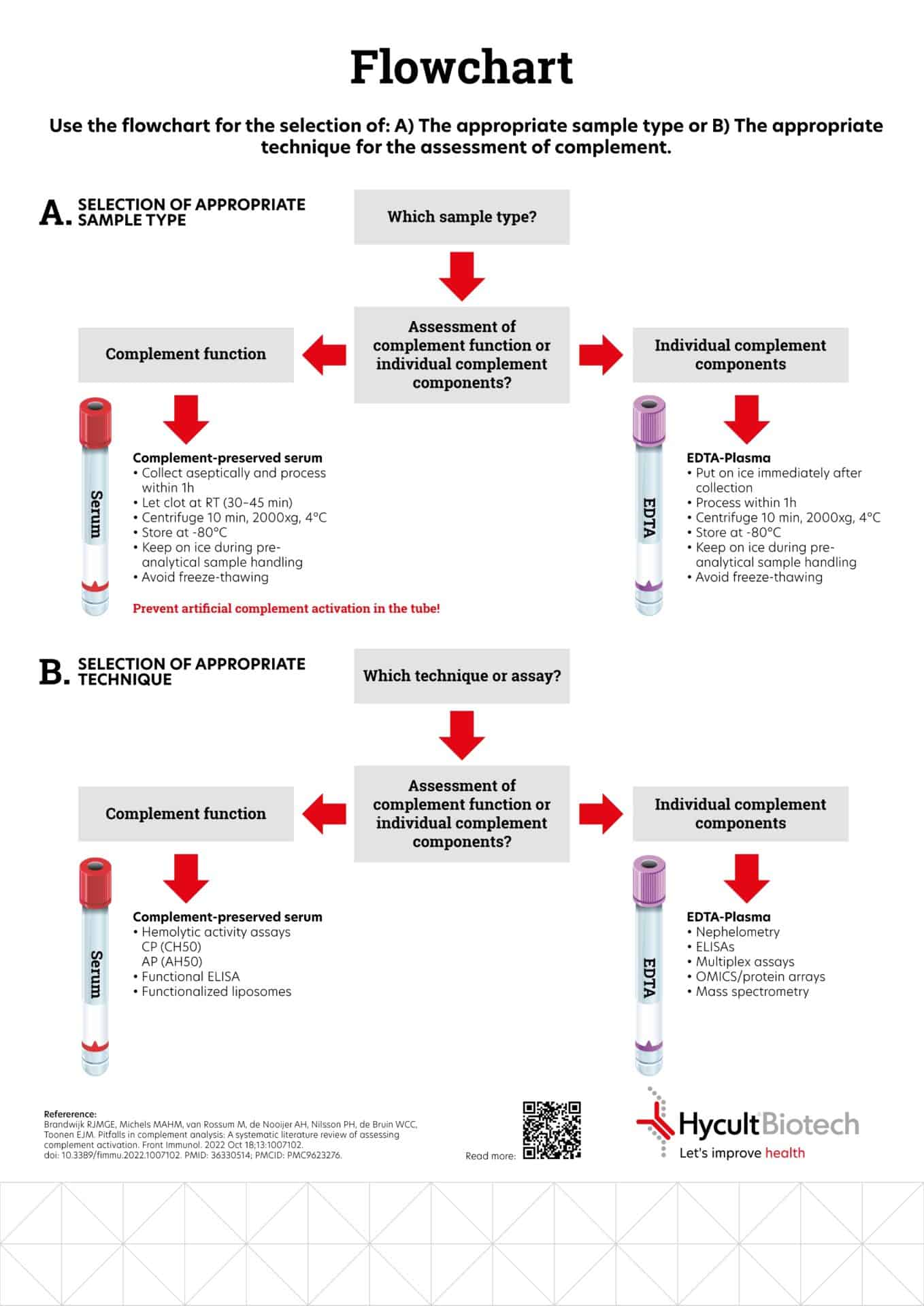
To choose the correct sample type, it is essential to begin by distinguishing whether you are assessing the overall function of the complement system or the specific components. When your focus is on assessing complement function, it is crucial to utilize complement-preserved serum. Conversely, when you intend to evaluate individual complement components, EDTA-plasma should be employed. The flowchart presented below assists you in determining the suitable sample type and guides you toward the most suitable sample pretreatment method.
Appropriate technique
If you wish to determine which technique or assay is best suited for your research, it is also essential to identify whether you are conducting an assessment of complement function or an assessment of individual complement components. When assessing complement function, the most suitable techniques include hemolytica activity assays, functional ELISA or functionalized liposomes. For the evaluation of individual complement components, nephelometry, ELISAs, Multiplex assays, OMICS/protein arrays, or mass spectrometry are the preferred methods.
You can find optional guidelines below or reach out to us directly to help you.
Refererence:
Brandwijk RJMGE, Michels MAHM, van Rossum M, de Nooijer AH, Nilsson PH, de Bruin WCC, Toonen EJM. Pitfalls in complement analysis: A systematic literature review of assessing complement activation. Front Immunol. 2022 Oct 18;13:1007102. doi: 10.3389/fimmu.2022.1007102. PMID: 36330514; PMCID: PMC9623276.
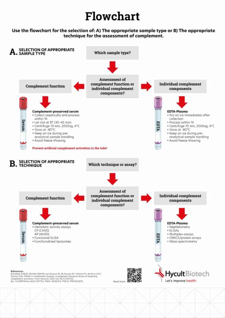
Flowchart
This chart helps you with the selection of the appropriate sample type and technique for complement analysis.
Use the flowchart for the selection of:
(A) the appropriate sample type or
(B) the appropriate technique for the assessment of complement.
Discover our range of solutions in C3
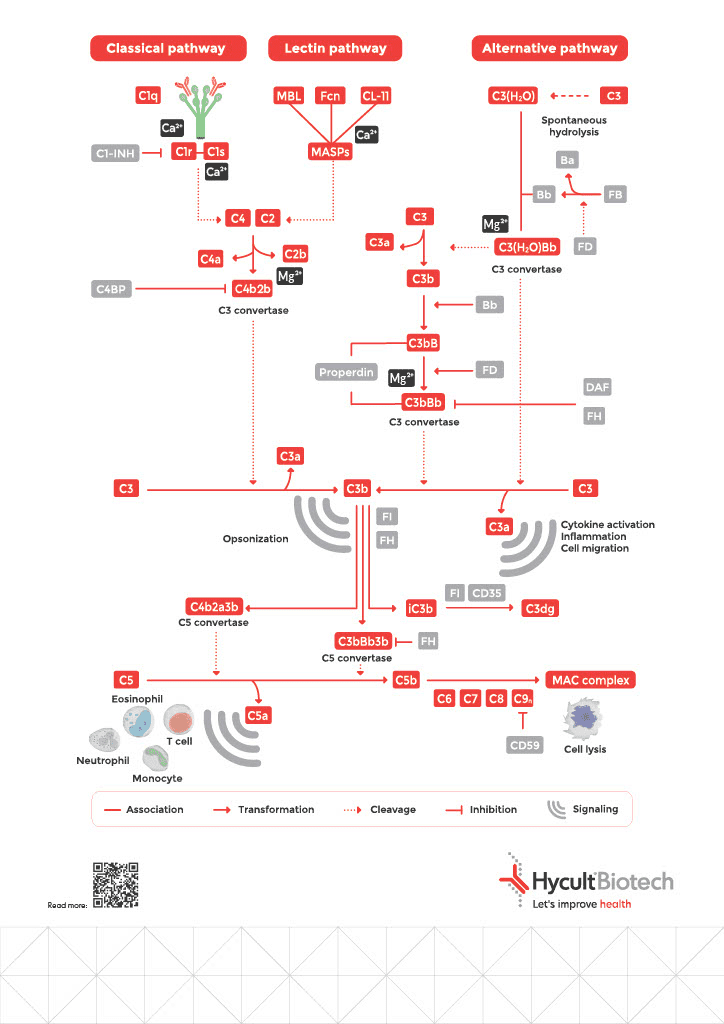
3 complement pathways
The complement system is a crucial part of the immune system, consisting of over 30 proteins that work together to help identify and destroy foreign pathogens. There are three major pathways of complement activation: the classical pathway, the lectin pathway, and the alternative pathway.
The classical pathway is activated when antibodies bind to specific antigens on the surface of pathogens. This binding triggers a cascade of complement proteins to bind and activate one another, ultimately leading to the formation of the membrane attack complex (MAC) that punctures and destroys the pathogen’s cell membrane.
The lectin pathway is activated when proteins called lectins bind to certain sugars on the surface of pathogens. This binding triggers a similar cascade of complement protein activation, leading to the formation of the MAC and pathogen destruction.
The alternative pathway is constantly active at low levels and can be triggered by the presence of certain molecules on the surface of pathogens or damaged host cells. This pathway also leads to the formation of the MAC and pathogen destruction.
All three complement pathways work together to provide a rapid and effective defense against invading pathogens. They also play a role in regulating the immune response and removing damaged host cells. Dysfunction of the complement system can lead to various diseases, including autoimmune disorders and infections.
We are glad to support you!
Take advantage of our dedicated support team for any technical assistance you need while using our products or considering them for your research needs.