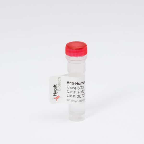
C3, Mouse, mAb 11H9
The monoclonal antibody 11H9 recognizes mouse complement component C3 and its activation products, C3b, iC3b, C3d and C3dg.
Read more€133.00 – €510.00
The monoclonal antibody 11H9 recognizes mouse complement component C3 and its activation products, C3b, iC3b, C3d and C3dg. The complement system is an important part of the humoral response in innate immunity, consisting of three different pathways. The third complement component, C3, is central to the classical, alternative and lectin pathways of complement activation. Activation products of the complement cascade contain neo-epitopes that are not present in the individual native components. The complement factor C3 consists of an alpha- and a beta-chain.
The synthesis of C3 is tissue-specific and is modulated in response to a variety of stimulatory agents. C3 is the most abundant protein of the complement system with serum protein levels of about 1.3 mg/ml. An inherited deficiency of C3 predisposes the person to frequent assaults of bacterial infections. In ulcerative colitis, and idiopathic chronic inflammatory bowel disease, the deposition of C3 in the diseased mucosa has been reported.
Proteolysis by certain enzymes results in the cleavage of C3 into C3a and C3b. C3b becomes attached to immune complexes and is further cleaved into iC3b, C3c, C3dg and C3f. The monoclonal antibody 11H9 recognizes both intact C3 and its cleaved products C3b, iC3b, C3d and C3dg. These activation products are present in acute as well as chronic inflammatory conditions. In chronic inflammatory condition, primarily the C3dg product resides at the place of inflammation (C3c being cleared) which is recognized by antibody 11H9. The chronic processing/activation of C3 is taking place at a lower level, which would reduce detection of the C3 fragments C3b, iC3b, and C3c.
W: A- reduced sample treatment and SDS-PAGE was used. The band sizes are ~110-115 kDa (;-chain), ~80 and 70 kDa (Ref.1).
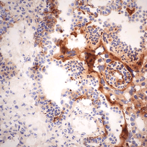
F:- Tissue sections were fixed in acetone. Antigen retrieval was performed using 50% formic acid for 5 min or microwaved (700 W) in antigen unmasking solution (Ref 1, 3).
IF: Fixation in 4% paraformaldehyde in PBS pH 7.4. Vibratome sections of 4 µm;m. Pretreated with 3% hydrogen peroxide for 20 min to quench endogenous peroxidases. Microwaved in antigen unmasking solution for 2-5 minutes as antigen retrieval. (Ref.3).
You may be interested in…
-
View product €1,784.00
-
View product €133.00 – €1,245.00
-
View product €133.00 – €456.00
