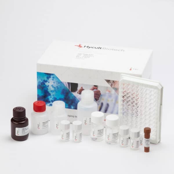
C4, Mouse, ELISA kit
€894.00 – €1,443.00
The complement system play important roles in both innate and adaptive immune response and can produce an inflammatory and protective reaction to challenges from pathogens before an adaptive response can occur. There are three pathways of complement activation. The classical pathway, the lectin pathway and the alternative pathway. The C3 and C5 convertases are enzymatic complexes that initiate and amplify the activity of the complement pathways and ultimately generate the cytolytic MAC.
The C4 glycoprotein is a large key molecule in the activation of the classical and lectin pathway. The formed proteolytic complexes of both pathways lead to cleavage of C4 thereby releasing the anaphylotoxin C4a and the pathway activating C4b. Binding of C4b to the cell surface leads to the formation of the C5 convertases, which is the start of the terminal pathway of complement. C4 functions as an acute phase protein and has a concentration of 250-500 ug/ml in healthy individuals. The majority of the protein is synthesized in the liver but also locally by among others monocytes, macrophages, lung, spleen, kidney and intestinal epithelial cells. Its expression is regulated by 4 genes, C4A and C4B, which are located in the HLA class III region. Although 99% identical, they have a different activity profile which is due to substrate specificity. C4A is more reactive and binds amino and thiol groups, while C4B binds more rapidly to hydroxyl groups. After activation of C4 the nascent C4b is quickly inactivated by its reaction with water. Only surface bound C4b forms the basis of formation of the CP/LP convertases. Total C4 concentration reflects potential to activate the complement system via the classical or lectin pathway.
Changes in complement levels may reflect chronic and/or recurring inflammation. C4 helps to prevent onset of autoimmune disease. Although rare, C4 deficiency is associated with SLE and chronic mucosal infections. Complement has a functional role in clearance of immune complexes or apoptotic cells.
Incomplete removal could lead to formation of autoantibodies.
Furthermore, complement is relevant in tolerance and deletion of autoreactive B cells.
The efficient format of a plate with twelve disposable 8-well strips allows free choice of batch size for the assay.
Samples and standards are incubated in microtiter wells coated with antibodies recognizing Mouse C4.
Peroxidase-conjugated antibody will bind to the captured C4.
Peroxidase-conjugate will react with the substrate, tetramethylbenzidine (TMB). The enzyme reaction is stopped by the addition of oxalic acid.
The absorbance at 450 nm is measured with a spectrophotometer. A standard curve is obtained by plotting the absorbance (linear) versus the corresponding concentrations of the Mouse C4 standards (log).
The Mouse C4 concentration of samples, which are run concurrently with the standards, can be determined from the standard curve.
