TLR9, Human, mAb 5G5
€133.00 – €368.00
The monoclonal antibody 5G5 recognizes human Toll-like receptor 9. Toll-like receptors (TLRs) are
highly conserved from Drosophila to humans and share structural and functional similarities. TLRs
constitute of a family of pattern recognition receptors (PRRs) that mediate cellular responses to a
large variety of pathogens (viruses, bacteria, and parasites) by specific recognition of so-called
‘pathogen-associated molecular patterns’. Activation of TLRs, a family of at least 11 differentmembers
that function either as homo- or heterodimers, leads to activation of NFκB-dependent and IFNregulatory
factor-dependent signaling pathways. TLRs have a central role in innate immunity and are
also required for the development of an adaptive immune response. TLRs are expressed by various
cells of the immune system, such as macrophages and dendritic cells. They recognize and respond to
molecules derived from bacterial, viral and fungal pathogens.
Whereas most TLRs are expressed on the cell surface, TLR9 is expressed intracellularly within one or
more endosomal compartments and recognizes nucleic acids. TLR9 detects a rather subtle difference
in the DNA of vertebrates compared with that of pathogens. Vertebrate genomic DNAs have mostly
methylated CpG dinucleotides where bacterial and viral DNAs have unmethylated CpG dinucleotides.
TLR9 undergoes relocation from endoplasmic reticulum to CpG-ODN-containing endosomes. In these
endosomes TLR9 becomes a functional receptor after proteolytic cleavage. TLR9 exists as a
preformed homodimer and CpG-ODN binding promotes its conformational change, bringing the
cytoplasmic TIR-like domains close to each other. This allows a recruitment of the key adapter protein
MyD88 which initiates a signalling cascade. The only human immune cell types known to
constitutively express TLR9 and to be activated by CpG ODN are pDCs and B cells. TLR9 triggering
induces an activation phenotype in the B cells and pDCs, characterized by the expression of
costimulatory molecules, resistance to apoptosis, and induces Th1-type immune response profiles.
IF: cells were fixed with 2% formalin for 15 minutes at RT and permeabilized with a mAb (4 µg/400 µl) containing buffer (PBS, 0.2% BSA, 0.2% saponin)- for 1 hour. (Ref.1)
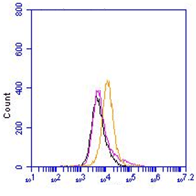
FC: RAW264.7 cells were fixed for 15 minutes with 4% formalin and permeabilized (PBS, 0.5%BSA, 0,5% saponin) at RT. (Ref.1)
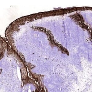
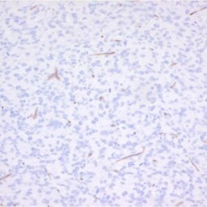
P: paraffin embedded tissues 5 µm;m sections were made. After antigen retrieval (0.01mol/l, pH6 sodium citrate) and quenching of endogenous peroxidase, sections were blocked with 0.5% ovalbumin and 0.1% gelatin for 20 minutes at RT. Sections were incubated with 5G5 for 1 hour at 37 °C. (Ref.3)
W: reduced lysates were resolved by 10% SDS-PAGE and blotted on nitrocellulose. After blocking with 5% skimmed milk TLR9 was detected with 2 µg/ml 5G5. (Ref.1)
