MBL-C, Mouse, mAb 14D12
€133.00 – €510.00
Mannose binding lectin (MBL), also called mannose- or mannan-binding protein (MBP), is a member of the group of collectins. MBL is an important pattern-recognition receptor in the innate immune system. The protein mediates innate immune responses, such as activation of the complement lectin pathway and phagocytosis, to help fight infections. MBL is an oligomeric lectin that recognizes carbohydrates as mannose and N-acetylglucosamine on pathogens. MBL contains a cysteine rich, a collagen like and a carbohydrate recognition domain. Binding of MBL leads to the activation of MBL-associated serine proteases (MASP’s). Activated MASP-2 cleaves C4 and C2 in a similar way as C1s do for the classical pathway (CP) leading to the formation of C4b2a, cleavage of the classical pathway convertase C3, and eventually complement activation up to the formation of the membrane attack complex. MBL is able to activate the complement pathway independent of the classical and alternative complement activation pathways.
MBL is predominantly synthesized by hepatocytes and has been isolated from the liver or serum of several vertebrate species. Only one form of human MBL has been characterized, while two forms are found in rhesus monkeys, rabbits, rats and mice. The mouse forms are known as MBL-A and MBL-C.
The MBL-C concentrations in serum are about 6-fold compared to that of MBL-A. MBL-A, but not MBL-C, was found to be an acute phase protein in casein and LPS-injection models. MBL-C exists in higher oligomeric forms than MBL-A. The monoclonal antibody 14D12 is a calcium-dependent antibody.
.
IA: Biotinylated 14D12 was used as detector in ELISA at 1 µg/ml TBS/Tween-20/Ca (Ref.1)
IF: Hyphae of C. albicans were fixed on poly-l-lysin coated slides with ice-cold aceton. Slides were incubated for 1h at RT with 14D12. (Ref.3)
W: On a 12% non-reducing SDS-PAGE a prominent band of ca. 50Kda is detected. Under reducing conditions a 26 KDa band is detected. (Ref.2)
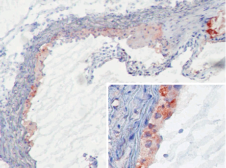
MBL-C (clone 14D12) deposition in developing murine atherosclerotic lesions following 10 weeks of high fat feeding. MBL-C was detected in and around invading macrophages invading the intima (insert). MBL-C bound, similar to MBL-A, at sites of necrosis (upper right corner). No MBL-C binding was shown in the media or on fibrous caps covering the thickened intima.


